Recent Press Releases
Video on the IVF projectTheMarker (Haaretz Journal), June 2016
|
 |
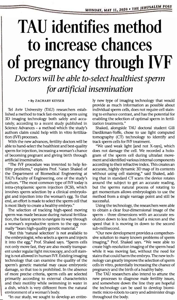
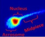
New microscopy may identify best sperm cellsTel Aviv University Newsroom, January 2016 “New microscopic technology from Tel Aviv University promises to be a game-changer in the field of reproductive assistance. A team of TAU scientists have devised a new method of microscopy allowing scientists to perform clinical sperm analysis without the use of staining, which can affect the viability of sperm samples. Sperm cells are nearly transparent under standard microscopy methods. Their optical properties differ only slightly from those of their surroundings, resulting in a weak image contrast. Sperm cells cannot be stained, if fertilization is the goal, because the process might damage the resulting fetuses. The challenge is to pinpoint strong sperm candidates without staining, while still being able to characterize their viability. The research, recently published in Fertility and Sterility, was led by Dr. Natan Shaked, PhD, of the Department of Biomedical Engineering at TAU’s Faculty of Engineering and his masters student, Dr. Miki Haifler, MD. Sperm cells for the study were obtained from the Male Fertility Clinic at Chaim Sheba Medical Center in Israel.” [Link]
|
 |
Nondestructive thickness measurements with subnanometer accuracy for fingureprint templatesLaser Focus World, November 2013 “Researchers at Tel Aviv University in Israel recently constructed compact, easy-to-align interferometric setups using common-path, low-coherence interferometry that is able to conduct nondestructive thickness measurements with subnanometer accuracy—for example, of fingerprint templates. Fingerprint templates created from transparent gels are a challenging type of sample for DH imaging due to their relatively deep and complex topography, which creates difficult phase-unwrapping problems. To overcome these problems, we have designed a low-cost, simplified, and noise-reduced low-coherence interferometric imaging system that can produce accurate depth profiles of fingerprints and be operated by inexpert users.” [Link]
|
 |
Optical microscopy quantifies live cells without labelsBiophotonics, October 2012 “Researchers at Tel Aviv University in Israel recently used IPM to record the phase of transparent samples without labeling at a rate of up to thousands of frames per second (a submillisecond rate). Professor Natan Shaked and colleagues in the department of biomedical engineering used live video to quantitatively image a monolayer of transparent HeLa human cervical cancer cells with subnanometer accuracy (albeit laterally diffraction-limited at a resolution of 600 nm) without labeling or physical contact with the sample.” The team demonstrated that optical thickness values of a cell can be used for quantitative microscopy to identify the various cell life-cycle stages and substages, growth rates, controlled or linear growth, and more – with no fluorescent dyes or special sample preparation.” [PDF] [Link]
|
 |
