New paper accepted to the Journal of Biophotonics
New paper accepted to the Journal of Biophotonics, 2015
Detection and controlled depletion of cancer cells using photothermal phase microscopy
Nir A. Turko, Itay Barnea, Omry Blum, Rafi Korenstein, and Natan T. Shaked
Abstract: We present a dual-modality technique based on wide-field photothermal (PT) interferometric phase imaging and simultaneous PT ablation to selectively deplete specific cell populations labelled by plasmonic nanoparticles. This combined technique utilizes the plasmonic reaction of gold nanoparticles under optical excitation to produce PT imaging contrast by inducing local phase changes when the excitation power is weak, or ablation of selected cells when increasing the excitation power. Controlling the entire process is carried out by dynamic quantitative phase imaging of all cells (labelled and unlabelled). We demonstrate our ability to detect and specifically ablate in vitro cancer cells over-expressing epidermal growth factor receptors (EGFRs), labelled with plasmonic nanoparticles, in the presence of either EGFR under-expressing cancer cells or white blood cells. The latter demonstration establishes an initial model for depletion of circulating tumour cells in blood. The proposed system is able to image in wide field the label-free quantitative phase profile together with the PT phase profile of the sample, and provides the ability of both detection and selective cell ablation in a controlled environment.
[Link]

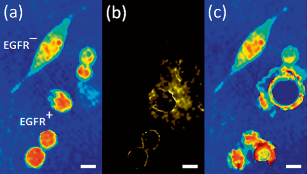
Quantitative phase imaging with molecular specificity and specific cell depletion. (a) Label-free quantitative phase profiles of mixed population of EGFR+/EGFR– cancer cells. (b) When weak modulated PT excitation is applied, selective phase contrast is generated in the modulation frequency only for the EGFR+ cancer cells labelled with plasmonic nanoparticles. (c) When stronger modulated PT excitation is applied, selective ablation of the EGFR+ cancer cells labelled with plasmonic nanoparticles occurs. White scalebars represent 10 µm upon sample. Figure is modified from DOI: 10.1002/jbio.201400095.