New paper accepted to Journal of Biophotonics
Yael Bishitz, Haniel Gabai, Pinhas Girshovitz, and Natan T. Shaked
Abstract: We propose to establish a cancer biomarker based on the unique optical-mechanical signatures of cancer cells measured in a noncontact, label-free manner by optical interferometry. Using wide-field interferometric phase microscopy (IPM), implemented by a portable, off-axis, common-path and low-coherence interferometric module, we quantitatively measured the time-dependent, nanometer-scale optical thickness fluctuation maps of live cells in vitro . We found that cancer cells fluctuate significantly more than healthy cells, and that metastatic cancer cells fluctuate significantly more than primary cancer cells. Atomic force microscopy (AFM) measurements validated the results. Our study shows the potential of IPM as a simple clinical tool for aiding in diagnosis and monitoring of cancer.

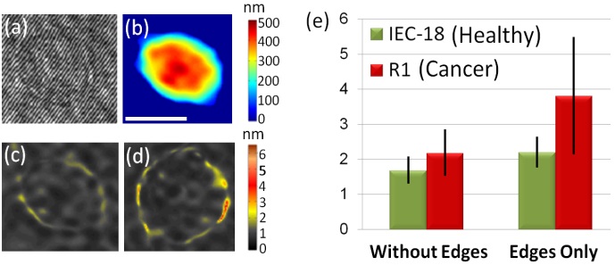
Wide-field IPM imaging of intestinal epithelial cells: (a) Off-axis interferogram of a healthy cell (IEC-18). (b) Quantitative optical thickness profiles of the healthy cell. (c) Fluctuation STD maps of the healthy cell. (d) Fluctuation STD maps of a cancer cell (R1). White scale-bar represents 10 µm. Colorbars represent optical thickness and optical thickness fluctuation STD in nanometers. (e) Comparison between the maximum fluctuation STD values of healthy cells (IEC-18) and cancer cells (R1) for the inside of the cell (without edges), and for the cell edges only. Cancer cells have been found to fluctuate significantly more than healthy cells. Figure is modified from the Journal of Biophotonics. Wiley 2013(c)