
In vitro viable model of the human placental barrier for transport studies
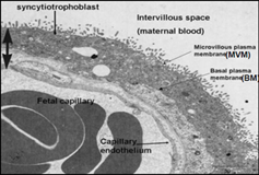
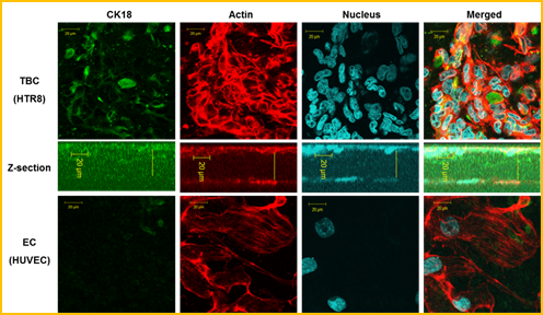
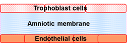
Maternal-to-fetal exchange of materials takes place at the placental barrier (PB), a multilayered structure that separates between maternal and fetal circulations. At the end of gestation the PB is composed of endothelial cells (EC) on the fetal side and the syncytiotrophoblast (STB) on the maternal side, which are separated by a basal lamina. In the first trimester it is composed of EC, basal lamina, cytotrophoblasts (CTB) and STB. It is widely accepted that "fetal programming" for health and disease in adult life takes place in utero. Huge efforts were also devoted to explore the normal and abnormal transplacental transport characteristics and mechanisms. Since in vivo studies in humans are limited due to accessibility and ethical issues, most of the knowledge was acquired via in vitro experiments, and mainly on the third trimester placenta. The ex vivo placental perfusion models have been regarded as the gold standard for studying placental transport. Regardless of their technical difficulties, their data represent only the global performance of the whole tissue of the PB. Many cellular models have been developed to simulate the PB structure, but most efforts were focused on the transport mechanisms across the maternal trophoblast cell (TC), while very little is known on the feto-placental endothelium. Recently, we constructed a viable tri-layer in vitro model of the human PB, which was made of a co-culture of human primary EC (HUVAC) and TC (HTR8) cell line on both sides of a denuded amniotic membrane (AM). Examination of TEM and immunofluorescent stained sections of the cellular structures of the PB model revealed the phenotype expressions of human EC and TC. This new approach enabled controlled in vitro experiments of transport and biological studies with laboratory setups that can mimic both the anatomical architecture of the PB and the in vivo environment of the placenta. Using similar culturing principles will enable future development of more advanced PBs that will represent the placenta of the 1st and 2nd trimesters, as well as pathological states.
 |  |
 |